📝 Introduction: The Quest for Faster, Clearer MRI Scans
The clarity and speed of magnetic resonance imaging (MRI) scans are paramount in medical diagnostics. However, traditional MRI methods are often slow and uncomfortable for patients. My colleagues, Praveen Kumar Pokala, Chandra Sekhar Seelamantula, and I have developed a novel approach that significantly accelerates MRI reconstruction while enhancing image quality. Our paper introduces the Generalized Fast Iteratively Reweighted Soft-Thresholding Algorithm (GFIRSTA), which optimizes complex-domain compressive sensing MRI (CS-MRI).
⚖️ The Challenge: Enhancing MRI Efficiency
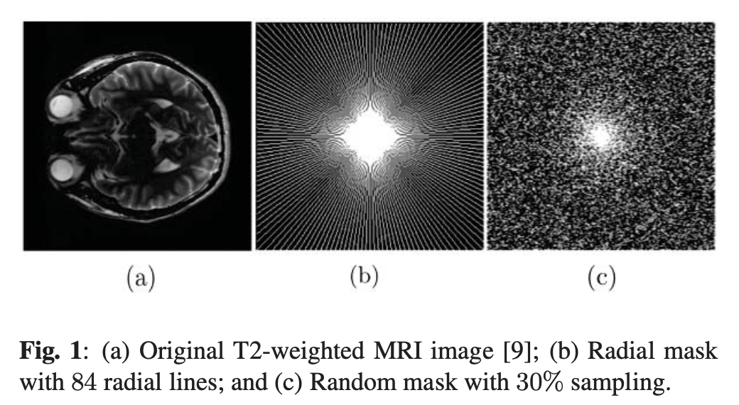
MRI techniques traditionally require long scan times to gather sufficient data for high-quality images, limiting their use in urgent care. Our research leverages compressive sensing (CS), a method that reconstructs images from undersampled data, to address this bottleneck effectively.
”In the end, we’ll all become stories.” - Margaret Atwood
🌱 Innovative Solution: GFIRSTA Explained
GFIRSTA uses a weighted ℓ1-minimization approach under tight frames, adapting weights iteratively for superior reconstruction. This method reduces artifacts typical in accelerated MRI techniques, paving the way for quicker and more accurate diagnostics.
🎨 Deep Dive: The Math Behind GFIRSTA
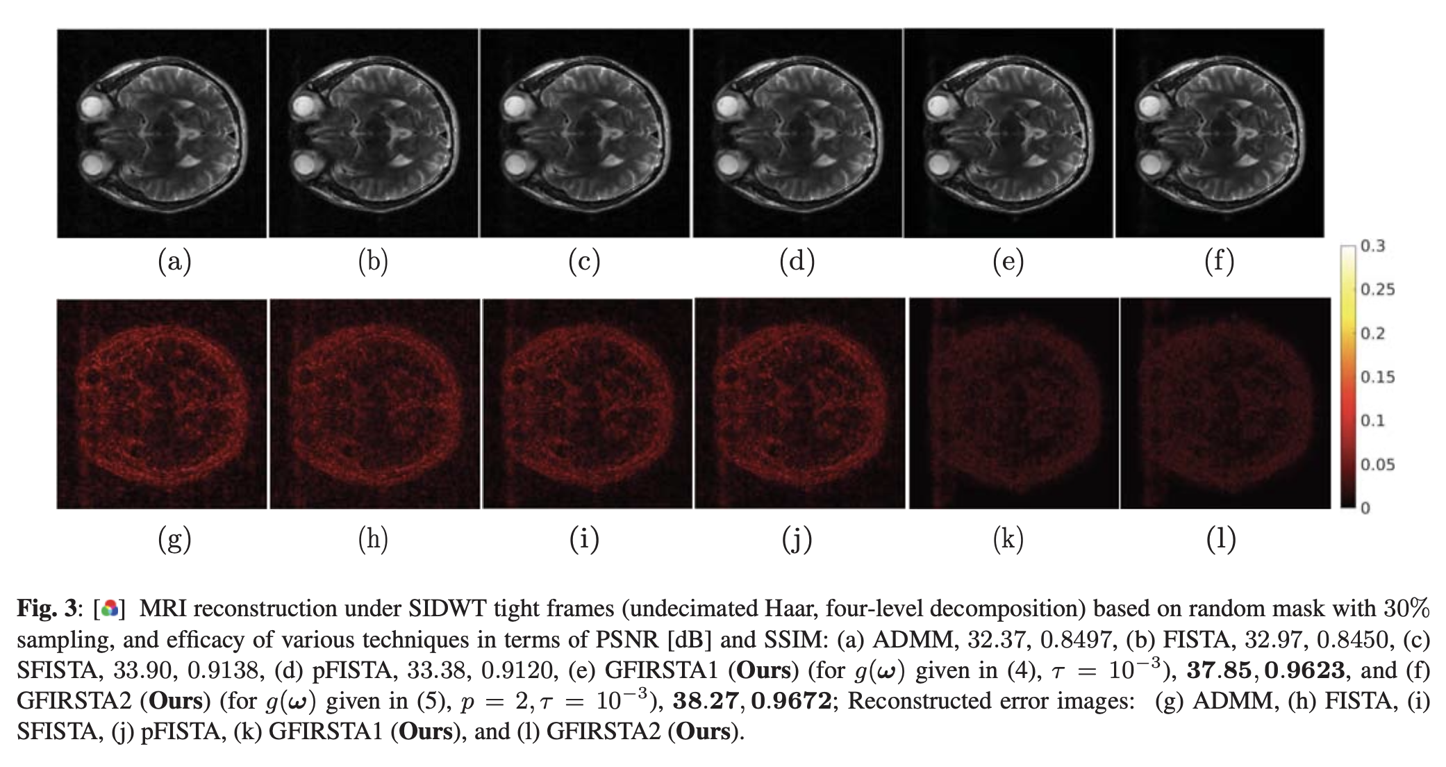
The core innovation of GFIRSTA lies in its approach to sparse coding in the complex domain under tight frames. Here is an in-depth explanation of the mathematics involved:
Sparse Coding and Tight Frames: Compressive sensing MRI involves solving an optimization problem to reconstruct an image from undersampled data. We express this as:
where 𝑥 is the image to be reconstructed, 𝐴 is the sensing matrix, 𝑦 is the sampled k-space data, and Ѱ is a sparsifying transform under tight frames, meaning Ѱ𝐻Ѱ = 𝐼, the identity matrix.
Weighted ℓ1-Minimization: Our approach extends traditional ℓ1-minimization by introducing a weight vector ω, which is updated iteratively:
where ⊙ denotes element-wise multiplication. The weighting adapts to emphasize less shrinking of large-valued coefficients, preserving important image details.
Wirtinger Calculus for Complex Optimization:
MRI data is complex, involving real and imaginary parts. Wirtinger calculus allows for the efficient handling of gradients in complex spaces:
where 𝚡 is the complex conjugate of 𝑥 and Re denotes the real part. This formula is used to compute the gradient of the loss function in our optimization.
Using this gradient, the update equation in our iterative algorithm becomes:
where η is the learning rate, reflecting how large each step is during the optimization.
🌟 Visual Evidence of Superiority
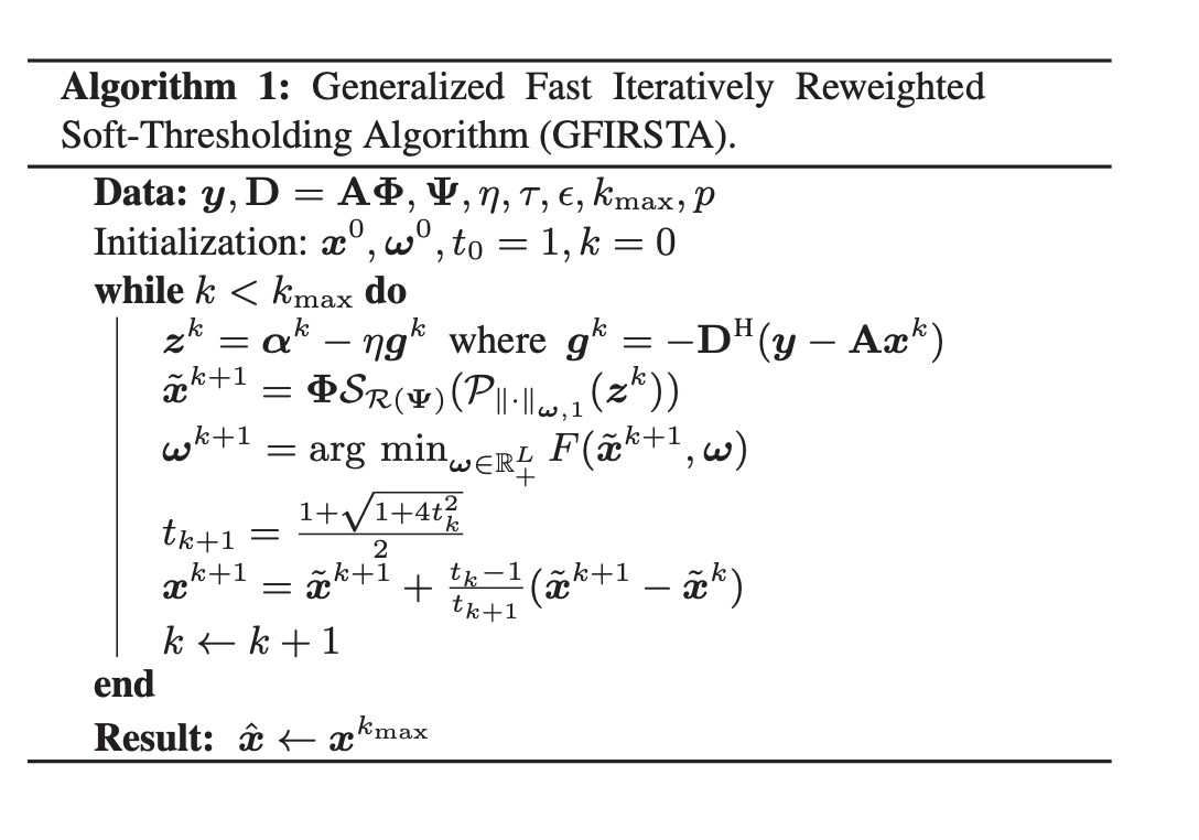
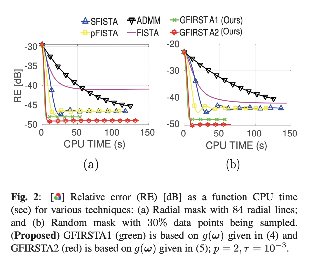
The effectiveness of GFIRSTA is demonstrated through extensive testing, where it consistently outperformed existing state-of-the-art techniques like FISTA and ADMM in terms of both the peak signal-to-noise ratio (PSNR) and structural similarity index metric (SSIM). Below are key visuals demonstrating these advancements:
Graph showing the rapid convergence and superior performance of GFIRSTA in MRI reconstruction.
📑 The Broader Impact
The implications of GFIRSTA extend beyond just faster scans. By improving diagnostic capabilities, it enhances patient outcomes in clinical settings and opens new possibilities for emergency diagnostics. Furthermore, GFIRSTA’s adaptability could revolutionize imaging techniques across various medical fields.
🎬 A Personal Journey: Behind the Scenes
This research journey has been both challenging and rewarding. Inspired by the potential to significantly impact patient care, we faced hurdles such as optimizing algorithm parameters and validating results under real-world conditions. These experiences have deepened my appreciation for the intersection of machine learning and medical imaging.
🗺️ Call to Action: Engage and Explore
I encourage you to read our full paper for a deeper dive into our methodologies and results. Please share this post with peers who might find it insightful, and do not hesitate to reach out with questions or comments. Let’s discuss how technology like GFIRSTA can continue to transform medical diagnostics.
💬 Concluding Thoughts
Our work on GFIRSTA represents a significant step forward in the field of medical imaging. By enhancing MRI technology, we’re not just improving a machine’s performance; we’re enabling quicker diagnoses, better patient care, and opening doors to new research opportunities.